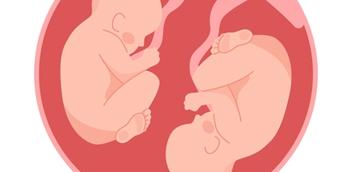
Kell sensitization can cause fetal anemia, too
Immunological reactions to the Kell antigen can cause as much damage to the unborn as Rh disease.
Understanding what causes Rh disease and developing an effective way to prevent the disorder is one of the genuine success stories of modern medicine. In fact, the administration of Rhesus immune globulin before and after delivery has saved countless lives and led to a major reduction in the incidence of Rhesus alloimmunization in pregnancy. Unfortunately, immune globulins to prevent maternal antibody formation to other red cells antigens have yet to be developed. A case in point is the Kell (K1) antigen, which has become a growing concern to clinicians as it, too, contributes to hemolytic disease of the fetus/newborn (HDFN).
In the mid 1960s, studies indicated that Kell antibodies occurred in 1.6 per 1,000 reproductive-aged women.1 In 1995, that number had doubled to 3.2 cases per 1,000 women-for reasons that are still unclear.2 And while countries like the Netherlands and Australia routinely administer Kell negative blood to female children and women of reproductive age when a transfusion is required, the United States is still a little behind the times. Here red blood cells are routinely cross-matched only for the ABO and RhD blood groups. It's no surprise then that two thirds of American women with Kell antibodies can trace their sensitization to a history of previous blood transfusion.3 In fact, in most large series, antibodies to the Kell antigen account for approximately 10% of fetuses with HDFN who undergo intrauterine transfusions.4
Understanding the basic immunopathology
Over 91% of Caucasians and 98% of African-Americans are Kell (K1) negative; these individuals are designated kk phenotype. Kell-positive individuals are designated either Kk or KK depending on their zygosity. In those with a Kell-positive blood type, 98% of Caucasians and virtually 100% of African-Americans are heterozygous. Serologic testing with anti-K and anti-k reagents can accurately determine zygosity in the case of a K1-positive father. Therefore, if 9% of male partners are Kell positive and the majority of these are heterozygous-which translates into a 50% risk for an affected fetus-the overall risk for an affected fetus in a Kell alloimmunized pregnancy, when the paternal Kell type and zygosity are unknown, is only 4.5%.
Most maternal RBC antibodies, including anti-D, cross the placenta and attach to fetal red cells that exhibit the specific antigen. Once these sensitized cells pass through the fetal spleen, they are filtered out of the circulating RBC pool and broken down by reticuloendothelial cells. The free hemoglobin that is released by this process is then converted to bilirubin. The fetal bone marrow responds to the resulting anemia by increasing the number of early red cell precursors (reticulocytes and erythroblasts) that are released into the fetal circulation.
The Kell antibody seems to cause fetal anemia through a two-prong attack on fetal red cells. Like anti-D, sensitized fetal red cells are sequestered in the fetal spleen. However, an additional mechanism involves suppression of red cell production so that there is an inadequate response to the fetal anemia. Laboratory studies have confirmed a direct effect of anti-Kell antibodies on developing red cells.6 In addition, blood from fetuses in Kell alloimmunized pregnancies contain fewer circulating reticulocytes and lower levels of serum bilirubin, when compared to fetuses with anti-D antibody induced HDFN.7
Newsletter
Get the latest clinical updates, case studies, and expert commentary in obstetric and gynecologic care. Sign up now to stay informed.


