Test your ob/gyn knowledge in our DailyDx.
Test your ob/gyn knowledge in our DailyDx.
Test your ob/gyn knowledge in our DailyDx.
Test your ob/gyn knowledge in our DailyDx.
Test your ob/gyn knowledge in our DailyDx.
Test your ob/gyn knowledge in our DailyDx.

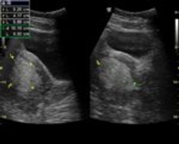
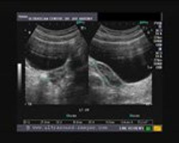
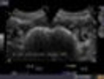
When a graafian follicle ruptures to release an oocyte, it is transformed into a corpus luteum. The corpus luteum is lined by a layer of granulose cells which rapidly become vascularized; some of these thin-walled vessels can rupture. This causes bleeding into the corpus luteum, resulting in the formation of a hemorrhagic cyst of the ovary.

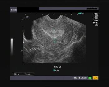
This case is of a young married female patient of 25 years age. She presented with bleeding per vagina and mild pain in the right side of the pelvis.
Test your ob/gyn knowledge in our DailyDx.
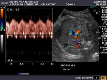
Significance of the umbilical cord: the umbilical cord can be aptly termed the life line for the fetus during its intrauterine life but can often be the cause for its misery in case of cord pathology.
What are echogenic intracardiac foci (EIF)? EIF are small, echogenic lesions seen (on sonography) inside the left or right ventricles of the fetal heart within the papillary muscles or chordae tendinae. These lesions are not attached to the wall of the ventricles.
Test your ob/gyn knowledge in our DailyDx.

Test your ob/gyn knowledge in our DailyDx.
Sending you an image of placenta, in late pregnancy (3rd trimester). I think it is low lying. Question is: can it be called mild previa?
Mother is sure of LMP 17th Nov/98 ( EDD = 24/8/99). On ultrasound scan (17/7/99) the average gestational age is only 31 weeks, ie the ultrasound derived EDD=18/9/99.
After delivery, there may be partial or complete retention of parts of the placenta or other decidual tissues within the uterus. This condition is termed “retained products of conception” (RPOC).
Test your ob/gyn knowledge in our DailyDx.
In this article month’s blog I’ll discuss one of the various uterine causes of infertility, focusing specifically on a condition in which the uterus is congenitally very small in size-the hypoplastic uterus.
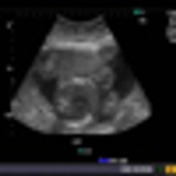
Normal 35 week pregnancy
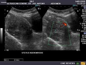
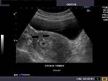
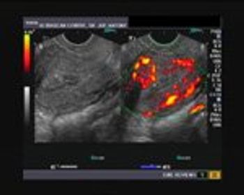
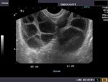
Is this lesion a fibroid or adenomyosis? Patient is a young (38n yr. old) married female with a history of dysmenorrhea and polymenorrhoea.
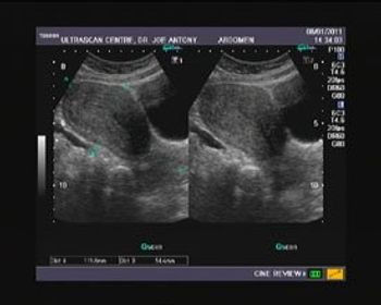
Images of "Bulky Uterus"
This young adult female (22 years age) had no significant complaints and was referred for a routine ultrasound scan of the abdomen to rule out any pathology. She complained of minor thyroid complaints, and ultrasonography suggested presence of Hashimoto’s thyroiditis in this lady.
Infertility is the inability to conceive despite frequent and unprotected intercourse between a man and his female partner for at least 1 year.